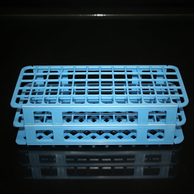
Technical Specs
Item
Test Tube Rack
Mounting Type
Rest on Table
Number of Compartments
60
Autoclavable
Yes
Material
Polypropylene
Color
Blue
Lab Rack Capacity
Holds 60 test tubes or conical tubes
Applications:
Suitable for product packaging and storage requirements in molecular biology and cell biology, laboratory medicine, genomics, proteomics, and other areas.
Product Features
The raw materials, premium polypropylene (PP, cap)/polyethylene (HDPE, bottle), have excellent physical and chemical indicators and very good pressure-resistance, impact-resistance and acid and alkali resistance.
PP materials can undergo autoclave sterilization at 121°C; HDPE can be stored at -80°C.
A full range of choices to meet different packaging and storage requirements.
Wash-free, no need for tedious pre-cleaning processing work, ready to use, greatly improves the packaging effect of users.
Thickened middle packaging to ensure transportation and storage safety.
Use of professional leak-proof design for bottle mouth, with no need for inner cover or inner gasket for protection, excellent sealability.
100% leak-proof to ensure safety even during air transportation; wide-mouthed design for easy access of liquid.
No biotoxicity, pyrogen-free, DNAse/RNAse-free, manufactured in a 100-thousand grade clean plant.
Sterilized using GAMMA-ray.
Introduction of imported digital-controlled production equipment with high precision
Fine details and comfortable touch
Bottle walls are even with high gloss, with no color difference; high uniformity between different batches.
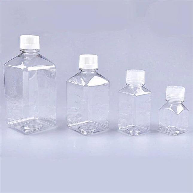
Features
Sterile
Bottle is used with PETG/PET
Screw cap is used with HDPE (High Density Polyethylene), free of heavy metals
Embossed and Graduated on two sides.
Shelf life: three year after month of production
All used materials meet the legal requirements of FDA
Temperature range from -60°C to 60°C
Non-autoclavability
Free of detectable DNase/RNase, human DNA and pyrogens
Features
Sterile
Bottle is used with PETG/PET
Screw cap is used with HDPE (High Density Polyethylene), free of heavy metal
Embossed and Graduated on two-sides
Shelf life: three year after month of production
All used materials meet the legal requirements of FDA
Temperature range from -60°C to 60°C
Non-autoclavability
Free of detectable DNase/RNase, human DNA and pyrogens
The PP bottles are for general laboratory use, ideal for lab specimens, chemicals and buffers.
Features:
Excellent chemical resistance to alcohols, alkalis and acids
Autoclavable
Can be stored in -40°C freezer
Durable
Leakproof design
Polypropylene caps included
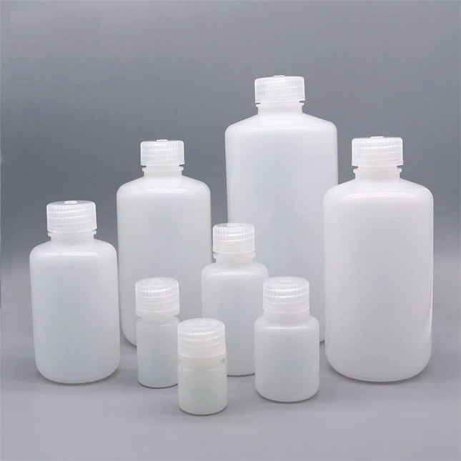
SKU
Capacity
Diameter x Height
Cap Size
BO3W10-10 BO3W10-100 BO3W10-1000
10 ml (0.3 fl oz)
23 x 48 mm
22 mm
BO3W15-10BO3W15-100BO3W15-1000
15 ml (0.5 fl oz)
27 x 48 mm
22 mm
BO3W30-10BO3W30-100BO3W30-800
30 ml (1.0 fl oz)
34 x 58 mm
31 mm
BO3W60-10BO3W60-100BO3W60-400
60 ml (2.0 fl oz)
40 x 82 mm
37 mm
BO3W125-10BO3W125-100BO3W125-250
125 ml (4.2 fl oz)
49 x 98 mm
43 mm
BO3W250-10BO3W250-150
250 ml (8.5 fl oz)
60 x 128 mm
43 mm
BO3W500-10BO3W500-120
500 ml (16.9 fl oz)
73 x 158 mm
56 mm
Applications:
Suitable for product packaging and storage requirements in molecular biology and cell biology, laboratory medicine, genomics and proteomics and other areas.
Product Features
The raw materials, premium polypropylene (PP)/polyethylene (HDPE), have excellent physical and chemical indicators, pressure-resistance, impact-resistance, and acid and alkali resistance.
PP materials can undergo autoclave sterilization at 121°C ; HDPE can be stored at -80°C.
Pre-washed/Ready-to-use: no need for tedious pre-cleaning
Thickened middle packaging to ensure transportation and storage safety.
Use of professional leak-proof design for bottle mouth, with no need for inner cover or inner gasket for protection, excellent sealability.
100% leak-proof to ensure safety even during air transportation; wide-mouthed design for easy access of liquid.
No biotoxicity, pyrogen-free, DNAse/RNAse-free, manufactured in a 100-thousand grade clean plant.
Sterilized using GAMMA-ray
Using automated production equipment, the bottles have fine details and evenly glossed walls. In addition, batches are incredibly uniform
By using high-grade molding and surface processing technologies, the bottles feature smooth interior and exterior surfaces that prevent the wicking effect of reagents, thereby significantly reducing sample wastage
Applications:
Suitable for product packaging and storage requirements in molecular biology and cell biology, laboratory medicine, genomics and proteomics and other areas.
Product Features
The raw materials, premium polypropylene (PP)/polyethylene (HDPE), have excellent physical and chemical indicators, pressure-resistance, impact-resistance, and acid and alkali resistance.
PP materials can undergo autoclave sterilization at 121°C; HDPE can be stored at -80°C.
Pre-washed/Ready-to-use: no need for tedious pre-cleaning
Thickened middle packaging to ensure transportation and storage safety.
Use of professional leak-proof design for bottle mouth, with no need for inner cover or inner gasket for protection, excellent sealability.
100% leak-proof to ensure safety even during air transportation; wide-mouthed design for easy access of liquid.
No biotoxicity, pyrogen-free, DNAse/RNAse-free, manufactured in a 100-thousand grade clean plant.
Sterilized using GAMMA-ray.
Using automated production equipment, the bottles have fine details and evenly glossed walls. In addition, batches are incredibly uniform.
By using high-grade molding and surface processing technologies, the bottles feature smooth interior and exterior surfaces that prevent the wicking effect of reagents, thereby significantly reducing sample wastage
SKU
Volume (ml)
Material
Color
Bottles per Bag
Total Bottles
336101
15
HDPE
Natural
20
400
337101
30
HDPE
Natural
10
200
338101
60
HDPE
Natural
10
200
339101
125
HDPE
Natural
10
100
340101
250
HDPE
Natural
10
100
341101
500
HDPE
Natural
5
50
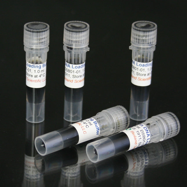
6X DNA Loading Buffer is a Ficoll 400-based ready-to-use DNA loading dye for tracking DNA migration during electrophoresis. it contains three dyes with different migration pattern: xylene cyanol FF migrates at approximately 4kb (Blue), bromophenol blue at 300bp (Purple) and orange G at 50bp (Orange) on a 1% agarose gel.
Storage: Room temperature or 4 °C
6X DNA Loading Buffer is a Ficoll 400-based ready-to-use DNA loading dye for tracking DNA migration during electrophoresis.
6X DNA Loading Buffer contains three dyes with different migration pattern: xylene cyanol FF migrates at approximately 4kb (Blue), bromophenol blue at 300bp (Purple) and orange G at 50bp (Orange) on a 1% agarose gel.
Storage: Room temperature or 4 °C
MSDS
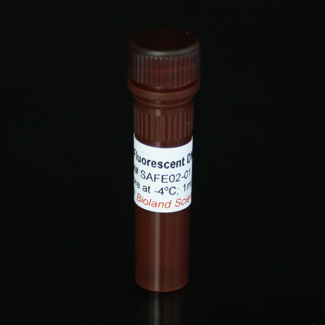
Ethium bromide replacement!
6xFluorescent DNA Loading Dye is formulated by mixing fluorescent dye with standard DNA loading dye and is used by adding to DNA samples directly. There is no need to add safe DNA gel stain or other fluorescent dye to the gel or running buffer. After the electrophoresis, the green DNA bands could be viewed under UV light or LED blue light.
Specifications
Storage
Store at 4 °C.
Shelf life
Two years from date of shipping.
Safety
Non-carcinogenic by the ames test. May cause skin and eye irritation. Always wear gloves when working with the product.
Disposal
Dispose as you would any other non-carcinogenic fluorescent dye (eg. Acridine orange; Propidium iodide).
Protocol
Prepare a 100 ml agarose or polyacrylamide solution with no stain. Mix gently to avoid bubbles.
For agarose gels, let the solution cool down to 60 - 70 oC before casting the gel. For polyacrylamide gel, add APS and TEMED and cast the gel according to regular protocol.
Mix samples or DNA marker with SafeRed dye at a 1:5 (dye : sample) dilution rate.
Following electrophoresis, view the results under UV and LED blue light.
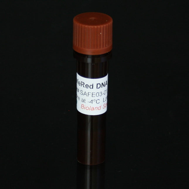
Ethium bromide replacement!
6x Red Fluorescent DNA Loading Dye is formulated by mixing fluorescent dye with standard DNA loading dye and is used by adding to DNA samples directly. There is no need to add safe DNA gel stain or other fluorescent dye to the gel or running buffer. After the electrophoresis, the red DNA bands could be viewed under UV light.
Specifications
Storage
Store at 4 °C.
Shelf life
Two years from date of shipping.
Safety
Non-carcinogenic by the ames test. May cause skin and eye irritation. Always wear gloves when working with the product.
Disposal
Dispose as you would any other non-carcinogenic fluorescent dye (eg. Acridine orange; Propidium iodide).
Protocol
Prepare a 100 ml agarose or polyacrylamide solution with no stain. Mix gently to avoid bubbles.
For agarose gels, let the solution cool down to 60 - 70 oC before casting the gel. For polyacrylamide gel, add APS and TEMED and cast the gel according to regular protocol.
Mix samples or DNA marker with SafeRed dye at a 1:5 (dye : sample) dilution rate.
Following electrophoresis, view the results under UV.